hotodynamic Damage and Bleaching
Dye Characteristics
Photodynamic damage depends on illumination intensity. At 4-10 msec time resolution, saturating a camera (10,000,000 electrons/pixel) with the dye fluorescence, using a 1:1 tandem lens, allowed averaging of about 256 trials, each of 1 sec, without significant photodamage. Bleaching proved very variable in cat visual cortex
The similarity between the corti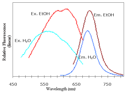 cal dye signal from a small population of neurons and an intracellular recording.
Two traces showing simultaneous intracellular and optical recording for 6 seconds, performed in deeply anesthetized cat: a condition in which spontaneous changes in membrane potential are highly synchronized in a large population of neurons. The intracellular recording is depicted by the green traces. The action potentials were truncated. The optical signal from the population next to the electrode is depicted by the red traces. Modified from Sterkin et al., 1998
Low biological noise probe for high-speed neural imaging of electrical signals
OI announces its new “blue” line of high-speed voltage sensitive dyes for cortical imaging, originally synthesized by Rina Hildesheim, and available exclusively from OI (patent pending; blue dye series includes RH-1691, and RH-1692). Voltage sensitive dyes (VSDs) are optical transducers of membrane potential changes. Applied to the brain, they bind to the external surface of the membranes of living cells without interrupting their normal function. Once introduced into a preparation, VSDs rapidly (within a microsecond) alter the intensity and/or wavelengths of fluorescent light they emit as a function of changes in neuronal membrane potential.