Doxorubicin(DOX)刺激引起的细胞内代谢变化
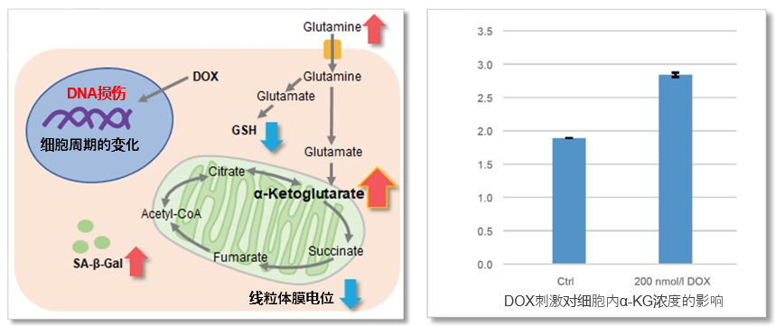
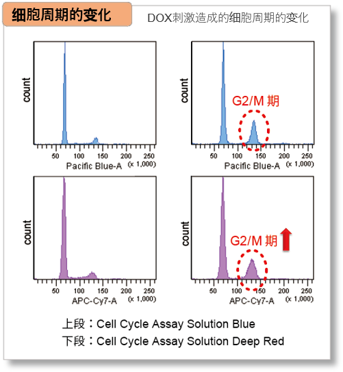
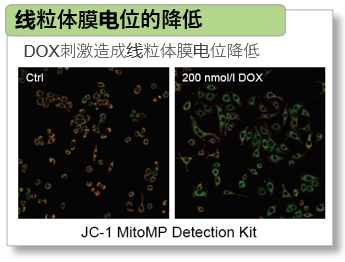
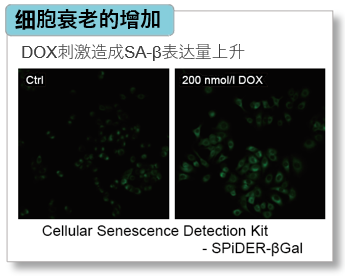
阿霉素(Doxorubicin, DOX)可以作用于 细胞周期的G2/M期,停止细胞的增殖并且细胞衰老,利用DOX作用于A549细胞,会导致胞内α-KG浓度增加。另外通过SG 03 Cellular Senescence Detection Kit – SPiDER-βGal检测细胞衰老、C548 Cell Cycle Assay Solution Deep Red / C549 Cell Cycle Assay Solution Blue检测细胞周期、MT09 JC-1 MitoMP Detection Kit检测线粒体膜电位的结果如下:
Sulfasalazine(SSZ)引起的细胞内代谢变化
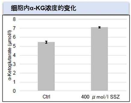
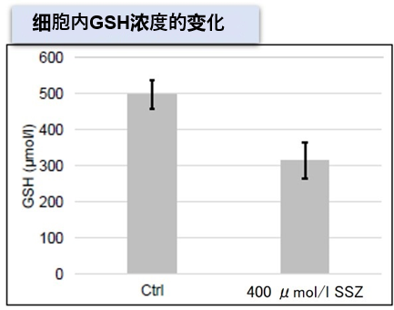
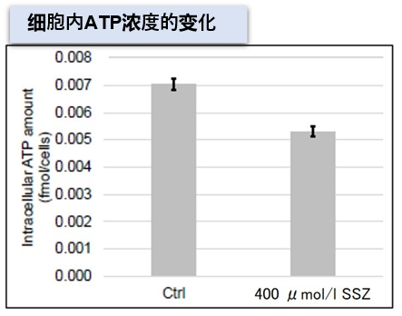
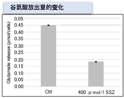
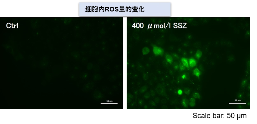
Sulfasalazine(SSZ)可以抑制细胞的胱氨酸/谷氨酸转运体(xCT)。用SSZ刺激A549细胞后,细胞内的α-KG、ATP、GSH、细胞放出的谷氨酸等变化用下列方法进行了检测。结果发现,SSZ刺激后细胞内的ATP、谷胱甘肽(GSH)、谷氨酸的放出量均减少,而细胞内的α-KG和ROS水平增加。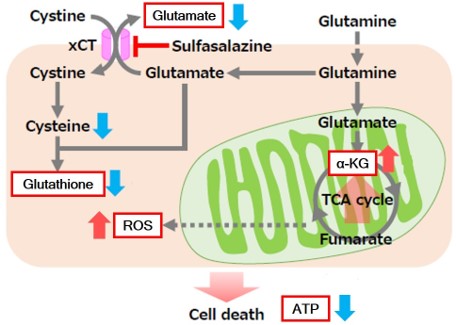
<使用产品>
・细胞内ATP:CK18 Cell Counting Kit-Luminescence
・细胞内GSH:G263 GSSG/GSH Quantification Kit II
・细胞内ROS:R252 ROS Assay Kit -Highly Sensitive DCFH-DA-
・胞外谷氨酸:G269 Glutamate Assay Kit-WST
<实验条件>
细胞:A549细胞(1 x 10⁶ cells) 暴露时间: 48 h
参考文献) Shogo Okazaki et al., “Glutaminolysis-related genes determine sensitivity to xCT-targeted therapy in head and neck squamous cell carcinoma”. Cancer Sci., 2019, doi:10.1111/cas.14182.
NASH诱导小鼠的肝脏组织的代谢变化
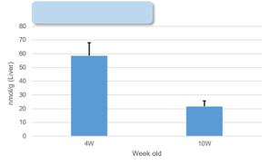
NASH(非酒精性脂肪肝)的病变组织中有ATP、α-KG、NAD的量减少的特点。使用4周龄的高脂肪食物投喂(引发NASH)的1型糖尿病模型小鼠(STAM模型)的肝脏组织,检测其中的ATP、α-KG、NAD水平的变化。结果显示,NASH诱导后10周龄的小鼠组中ATP、α-KG、NAD的浓度降低。

※详细的实验步骤请参考FAQ“是否有检测组织的实验例
<使用产品>
・组织内ATP:CK18 Cell Counting Kit-Luminescence
・组织内NAD:N509 NAD/NADH Assay Kit-WST
<实验参考文献>
| ATP |
Francesco Bellanti, et al., “Synergistic interaction of fatty acids and oxysterols impairs mitochondrial function and limits liver adaptation during nafld progression”, Redox Biology, 2018, 15, 86-96. |
| α-KG |
Jianjian Zhao, et al., “The mechanism and role of intracellular α-ketoglutarate reduction in hepatic stellate cell activation”, Bioscience Reports, 2020, 40, (3). |
| Ali Canbay, et al., “L‑Ornithine L‑Aspartate (LOLA) as a Novel Approach for Therapy of Non‑alcoholic Fatty Liver Disease”, Drugs, 2019, 79, 39-44. |
| NAD |
Jinhan He, et al., “Activation of the Aryl Hydrocarbon Receptor Sensitizes Mice to Nonalcoholic Steatohepatitis by Deactivating Mitochondrial Sirtuin Deacetylase Sirt3”, Mol. and Cell. Biol., 2013, 33, (10), 2047-55.
|

