| 产品编号 | 产品名称 | 产品规格 | 产品等级 | 产品价格 |
| 019-19741 | Anti Iba1, Rabbit(for Immunocytochemistry) 抗Iba1,兔(免疫细胞化学) |
50 μg | 免疫化学 | - |
| 016-20001 | Anti Iba1, Rabbit (for Western blotting) 抗Iba1,兔(用于免疫印迹) |
50 μg | 免疫化学 | - |
- 产品特性
- 相关资料
- Q&A
- 参考文献
 小胶质细胞特异性抗体
兔源Iba1抗体,无标签
Iba 1是在巨噬细胞/小胶质细胞中特异性表达的分子量为17,000的钙结合蛋白。近来小胶质细胞备受关注,除了在神经营养/神经保护中起作用外,也已被证实通过产生NO,TNF-α和IL-1β对神经造成损伤。
该产品是与小胶质细胞特异性反应的兔多克隆抗体,适用于与星形胶质细胞特异性抗GFAP单克隆抗体进行双染色。
◆特点
● 抗原:对应于Iba1的C末端的合成肽
● 形式:TBS溶液(1mg / mL)
● 纯化:兔抗血清的抗原亲和层析纯化
● 特异性:
对小胶质细胞和巨噬细胞具有特异性,但不与神经元和星形胶质细胞发生交叉反应。
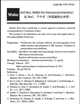
与人类,小鼠大鼠Iba1反应。
● 用法:
1.抗Iba1,兔(免疫细胞化学)适用于免疫细胞化学。 1-2μg/ mL使用
2.抗Iba1,兔(用于Westernblotting)适合于Westernblotting(免疫印迹)。 0.5-1μg/ mL使用。
◆应用
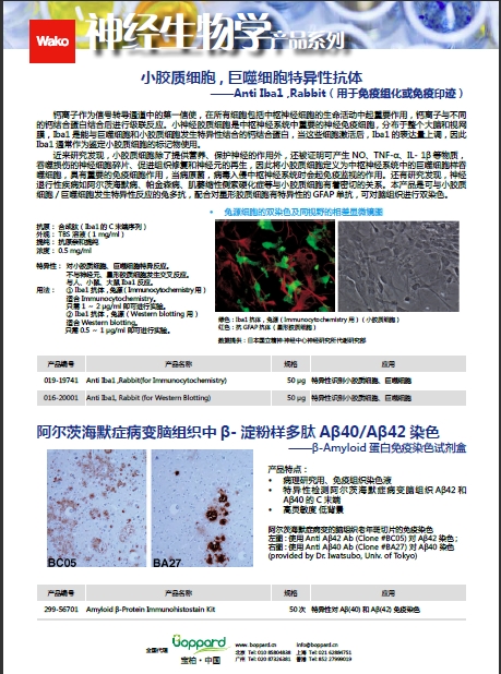
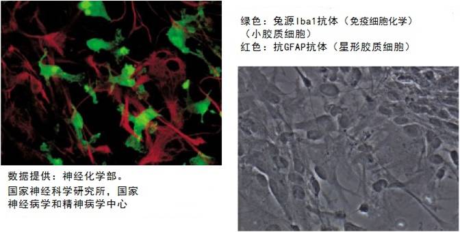
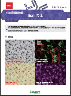
大鼠原代混合培养细胞双染色和相同视野的相位图像
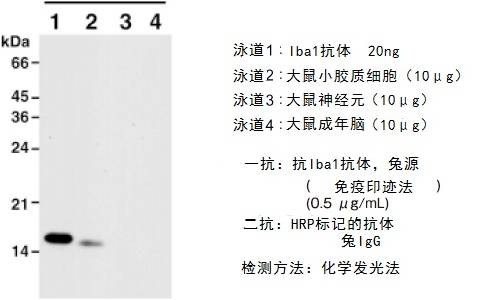
◆免疫印迹实验
◆推荐准备工作
a)石蜡切片
1. 脱蜡
100%二甲苯10分钟,2次
⇓
100%乙醇5分钟,2次
⇓
80%乙醇5分钟
⇓
70%乙醇5分钟
⇓
dH2O,5分钟,2次
2. 用0.01M PBS洗涤,2次
3. 在0.3%H 2 O 2的甲醇溶液中孵育30分钟
4. 用0.01M PBS洗涤,3次
5. 室温(RT)下用封闭缓冲液(1.5%正常山羊血清和0.001M PBS的1%BSA)孵育2小时。
6. 4℃下在封闭缓冲液中与抗Iba1抗体(0.5μg/ mL)(Wako目录号019-19741)孵育过夜。
7. 用0.01M PBS洗涤,3次。
8. 与生物素化的抗兔IgG抗体(1:200)在封闭缓冲液中室温孵育1小时。
9. 用0.01M PBS洗涤,3次。
10. 室温下与Elite ABC试剂(0.01M PBS中的试剂A(1:50)和试剂B(1:50))孵育1小时。
11. 用0.01M PBS洗涤,3次
12. 在过氧化物酶溶液中孵育(0.05M Tris缓冲液的0.01%过氧化氢和0.05%DAB)
◆实验步骤链接
b)冰冻切片
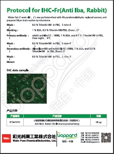
冷冻的小鼠脑组织或培养的细胞应用4%多聚甲醛-PBS灌注固定。然后制备组织切片。制备后,将组织切片和培养的细胞用抗Iba1抗体免疫染色。
欲了解相关产品信息请点击文字:
兔源Iba1抗体,有标签
鼠源Iba1抗体,无标签,单克隆抗体(NCNP24)
欲了解相关知识请点击文字:
巨噬细胞/小胶质细胞特异性蛋白抗体Iba1的应用
最新小胶质细胞研究动向与新型Iba1标签抗体
欲了解相关资料请点击文字:
Wako神经生物学抗体清单
巨噬细胞/小胶质细胞Iba1抗体
 |
 |
|
|
|
 |
 |
|
|
|
|
|
 |
|
β-Amyloid ELISA |
Protocol for IHC-Fr(Anti Iba Rabbit) |
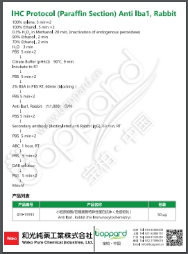
Protocol for IHC-P(Anti Iba Rabbit)
参考文献
|
产品编号 |
产品名称 |
2017年发表文献引用 |
|
|
019-19741 |
Anti Iba1, Rabbit (for Immunocytochemistry) |
[1] |
Tomov N, Surchev L. Punctate Staining as Indirect Evidence for Microglial Ramification[J]. Acta morphologica et anthropologica, 24: 1-2.<链接> |
|
[2] |
Barua S, Chung J I, Kim A Y, et al. Jak kinase 3 signaling in microgliogenesis from the spinal nestin+ progenitors in both development and response to injury[J]. Neuroreport, 2017, 28(14): 929.<链接> |
||
|
[3] |
Dilution A S. Supplementary Table S2. Antibodies used in immunohistochemical, flatmount and immunoblotting studies[J]. <链接> |
||
|
[4] |
Su W S, Wu C H, Chen S F, et al. Low-intensity pulsed ultrasound improves behavioral and histological outcomes after experimental traumatic brain injury[J]. Scientific Reports, 2017, 7(1): 15524. <链接> |
||
|
[5] |
Ebneter A, Kokona D, Jovanovic J, et al. Dramatic Effect of Oral CSF-1R Kinase Inhibitor on Retinal Microglia Revealed by In Vivo Scanning Laser Ophthalmoscopy[J]. Translational vision science & technology, 2017, 6(2): 10-10.<链接> |
||
|
016-20001 |
Anti Iba1, Rabbit (for Western Blotting) |
[1] |
Zhang X, Wang D, Pan H, et al. Enhanced expression of markers for astrocytes in the brain of a line of GFAP-TK transgenic mice[J]. Frontiers in neuroscience, 2017, 11. <链接> |
|
[2] |
Barua S, Chung J I, Kim A Y, et al. Jak kinase 3 signaling in microgliogenesis from the spinal nestin+ progenitors in both development and response to injury[J]. Neuroreport, 2017, 28(14): 929.<链接> |
||
|
[3] |
Peters D G, Purnell C J, Haaf M P, et al. Dietary lipophilic iron accelerates regional brain iron-load in C57BL6 mice[J]. Brain Structure and Function, 2017: 1-18.<链接> |
||
|
[4] |
Edwards A, Szklarczyk A, Ottenheimer D, et al. Matrix metalloproteinase activity stimulates N-cadherin shedding and the soluble N-cadherin ectodomain promotes classical microglial activation[J]. Journal of neuroinflammation, 2017, 14(1): 56.<链接> |
||
|
[5] |
Roche S L, Ruiz‐Lopez A M, Moloney J N, et al. Microglial‐induced Müller cell gliosis is attenuated by progesterone in a mouse model of retinitis pigmentosa[J]. Glia, 2017. <链接> |
||
jQuery(“.nltabbox”).slide({effect:”fade”});

